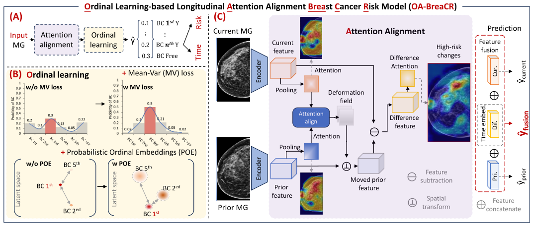
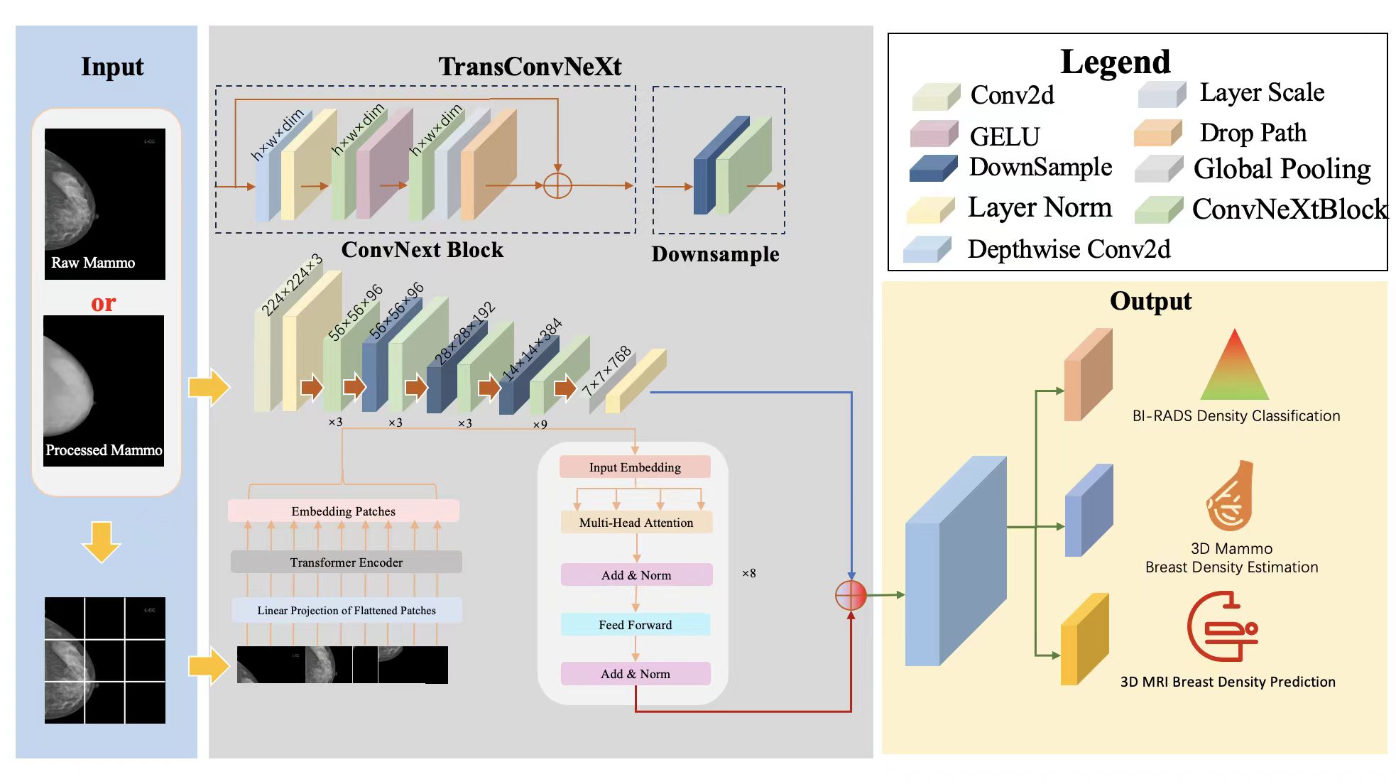
Multi-tasking Breast Density Classification
Project Leaders
Xinye Wang
Tianyu Xie
Partner Organisations
江苏集萃苏科思科技有限公司
Project Leaders
Xinye Wang
Tianyu Xie
Partner Organisations
江苏集萃苏科思科技有限公司
Project Example
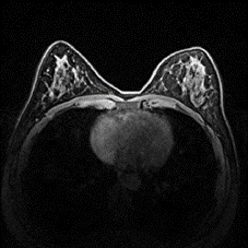
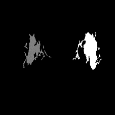
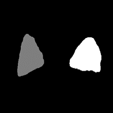
The first image is the MRI original image of the breast, the second image is the segmentation of dense tissue within the breast, and the third image is the segmentation of the entire breast
Project Leaders
Jinghong Song