APTw imaging and segmentation

Project Leaders

Qiuhui Yang

Partner Organisations

National Clinical Research Center for Cancer

Cancer Hospital & Shenzhen Hospital
APT fast imaging

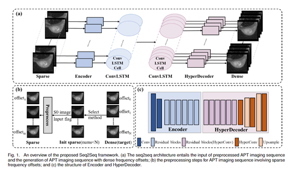
Reducing the acquisition time of the Amide Proton Transfer (APT) technique is crucial for its widespread application. One way to reduce the scanning time is by decreasing the frequency offsets used during image acquisition, which might compromise the quantification of the APT effect. In our study, we develop a deep learning-based model that can reconstruct dense frequency offsets from sparse ones, potentially reducing scanning time. We propose using convLSTM to extract both short and long-range spatial and frequency features of APT imaging sequences. Our proposed model outperforms other seq2seq models, achieving superior reconstruction with a peak signal-to-noise ratio of 45.8 (95% confidence interval (CI)): [44.9 46.7]), and a structural similarity index of 0.989 (95% CI:[0.987 0.993]) for the tumor region. We have integrated a weighted layer into our model to evaluate the impact of individual frequency offsets on the reconstruction process. The weights assigned to the frequency offset at ± 6.5 ppm, 0 ppm, and 3.5 ppm demonstrate higher significance as learned by the model. Experimental results demonstrate that our proposed model effectively reconstructs dense APT imaging sequences (+7 to -7 with 0.5 ppm as interval) from data with 21 frequency offsets, reducing scanning time by 25%. This work presents a method for lowering the APT imaging time, offering potential guidance for parameter settings in physical devices during APT imaging and serving as a valuable reference for clinicians.

Analysis of the entire group

Project Leaders

Qiuhui Yang

Partner Organisations

National Clinical Research Center for Cancer

Cancer Hospital & Shenzhen Hospital

APT Segmentation

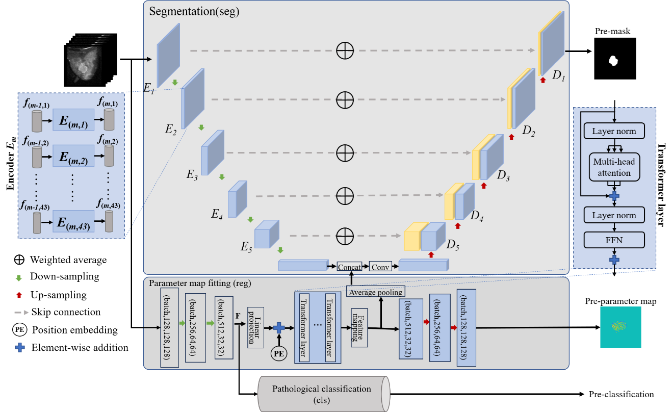
As a new magnetic resonance imaging technology with a unique contrast mechanism, Amide Proton Transfer Weighted (APTw) imaging has shown its potential in the diagnosis, treatment evaluation, and prognosis prediction of breast cancer. The signal contrast between the lesion and the surrounding glandular tissue is low on the anatomical images obtained using the APTw imaging sequence in comparison to conventional anatomical dynamic contrast-enhanced MR images, which leads to subjective and inconsistent identification of the lesion for clinicians. However, breast lesion regions and their surrounding glandular tissues exhibit different APT effects, which can be utilized to enhance the segmentation accuracy of breast lesion regions on acquired anatomical images of the APTw sequence. Therefore, this paper proposes a breast lesion region segmentation network based on the model that incorporates APTw parameter map fitting and pathological classification for automatic lesion region segmentation. In addition, this paper demonstrates the importance of each frequency in APTw imaging for clinical tasks. It improves the interpretability of the network, allowing clinicians to understand its functionality better. We conducted experiments on 164 cases of originally acquired images of the APTw sequence, with lesion regions being jointly labeled as ground truth by three senior radiologists. The results show that the proposed method performs well in lesion region segmentation on images of APT sequence. Compared with these advanced methods such as U-Net, SAM, and TransBTS, our method achieves higher accuracy. Additionally, the model's interpretable contribution to different frequency offsets aligns with clinical observations.

UniMRISegNet: Universal 3D Network for Various Organs and Cancers Segmentation on Multi-Sequence MRI

Project Leaders

Qiuhui Yang

Partner Organisations

National Clinical Research Center for Cancer

Cancer Hospital & Shenzhen Hospital
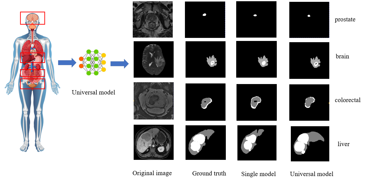
Magnetic Resonance Imaging (MRI) stands as a cornerstone in modern medical diagnostics, renowned for its unparalleled ability to provide exquisite soft tissue contrast. This advanced imaging modality has become an indispensable tool in patient screening processes. The versatility of MRI is underscored by its array of imaging sequences, including T1-weighted (T1w), T1-weighted contrast-enhanced (T1c), T2-weighted (T2W), T2-fluid attenuated inversion recovery (T2f), and Apparent Diffusion Coefficient (ADC), among others. Each sequence offers a unique perspective on the lesions within the body, providing crucial information that aids clinicians in formulating accurate diagnoses and treatment plans.

Despite its numerous advantages, the diversity of MRI sequences poses a substantial challenge for radiologists. They are often burdened with the arduous and time-intensive task of manually labeling multiple organs or cancerous regions in three-dimensional (3D) space, based on various MRI sequences. This process is not only labor-intensive but also prone to human error, which can impact patient care. Furthermore, existing automated segmentation methods are largely tailored to specific MRI sequences, organs, or types of cancer. This specialization results in a lack of generality, making these methods less adaptable to a broader range of medical imaging tasks. As the number of tasks for which these methods are trained increases, inference speeds tend to slow down, and the parameter counts, leading to increased computational complexity.

In light of these challenges, there is an urgent demand for the development of an automatic universal 3D segmentation network. Such a network would need to be capable of handling multiple MRI sequences, as well as segmenting various organs and types of cancer. This innovative approach would not only streamline the diagnostic process but also enhance the overall efficiency and accuracy of medical imaging analysis, ultimately contributing to improved patient outcomes.


MR image denoising

Project Leaders

Zhuoneng Zhang

Partner Organisations

National Clinical Research Center for Cancer

Cancer Hospital & Shenzhen Hospital
Magnetic resonance (MR) is a safe and radiation-free imaging technique used on living subjects, and the images produced by this technique are called MR images. These images enable physicians to diagnose patients accurately and quickly. However, noise caused by mechanical and environmental factors during MR image acquisition affects the quality of MR images, so it is important to remove the noise from MR images to improve the image quality.

MR is a technique that uses signals generated by atomic nuclei resonating within a magnetic field to be reconstructed for imaging. Atomic nuclei in human tissues (containing the base proton or neutron) magnetized in a strong magnetic field, the gradient field to give spatial positioning, radio frequency pulses to stimulate a specific frequency of the hydrogen proton into resonance, accept the excitation of the hydrogen proton chiropteric process releases energy, that is, the magnetic resonance signal, the computer will be the MR signals are collected, according to the intensity of the conversion into black and white grayscale, the composition of two-dimensional or three-dimensional form according to the position, and ultimately the composition of the MR image. . In the process of MRI, different images can be obtained by changing the influencing factors of the MR signal, and these different images are called sequences. Existing filtering methods, transform domain methods, statistical methods, and convolutional neural network (CNN) methods all aim to denoise a single sequence of an image without considering the relationship between multiple different sequences. They do not balance the extraction of high and low dimensional features in MR images and struggle to maintain a good balance between preserving image texture details and denoising intensity.

To overcome these challenges, this work proposes a controlled multimodal cross-global learnable attention network (MMCLANet) for MR image denoising. Our approach preprocesses images into multimodal connections and then uses a cross-global learnable attention network to extract and fuse image features between different modalities and within the same modality. In this process, a controlled noise level map is introduced to flexibly control the denoising intensity. In addition, we introduce a stochastic modality missing mechanism during model training, which can also effectively remove noise from MR images with missing modalities

Our proposed method outperforms the baseline method and other methods used in this study in terms of SSIM and PSNR. Our model also requires fewer FLOPS than the baseline method, which indicates better computational performance of our method.

We propose a model for multimodal denoising by exploiting the correlation between different sequences (i.e., different modalities) of MR images, which can handle both missing-modality and all-modality denoising tasks, improving the generalization and denoising performance of the model, and which employs a cross-global learnable attention network for multimodal fusion. The method can provide a reference idea for other medical image denoising of the same type.