Our Approach
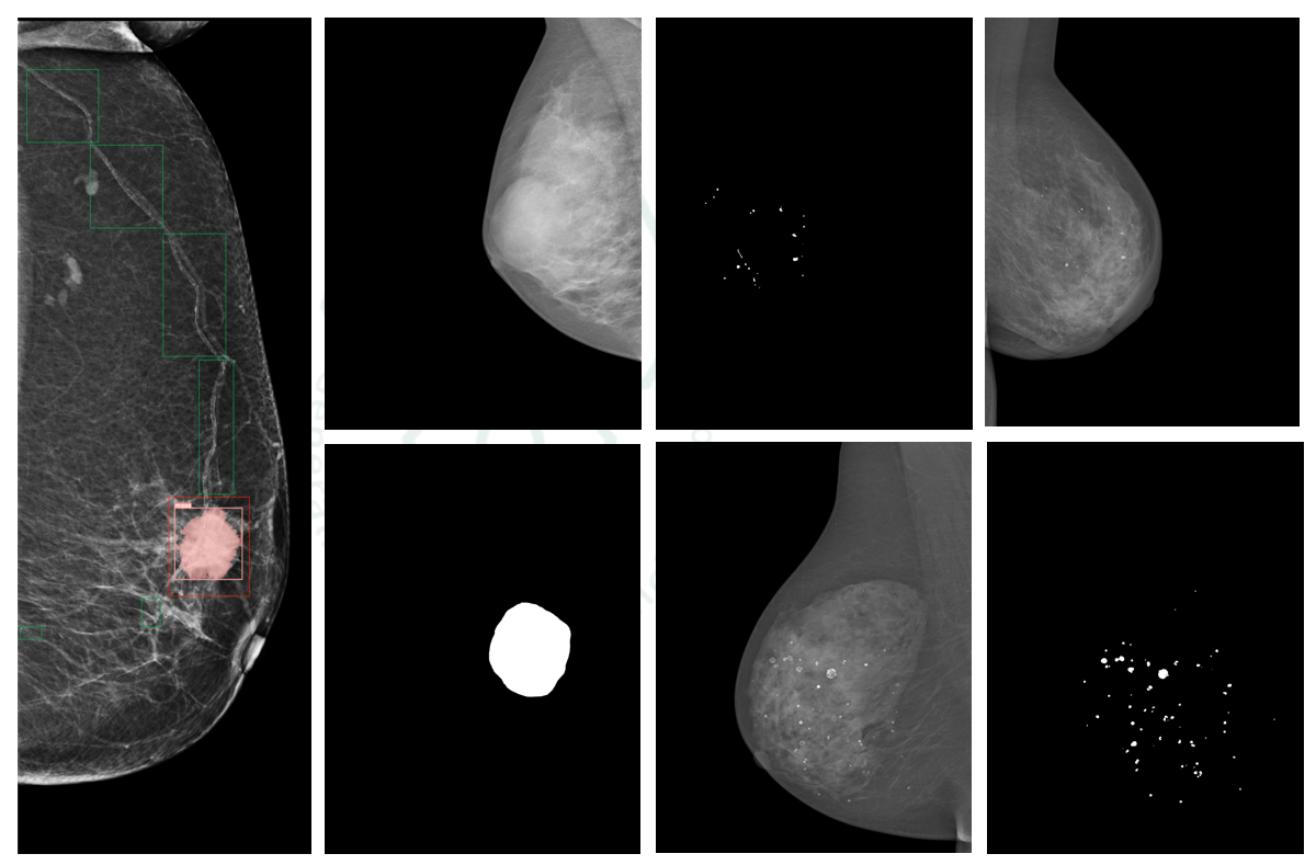
We have adapted the widely acclaimed U-Net architecture specifically for detailed multi-class segmentation of mammographic images. Our solution focuses on enhancing the detection and classification of calcifications and masses—critical indicators of potential malignancies whose precise identification is essential for effective treatment planning.
Key Innovations:
1.Multi-class Pretraining:** We leverage extensive multi-class pretraining to equip our model with a comprehensive understanding of diverse image attributes and abnormalities. This enables more nuanced and accurate image segmentation across various tissue types and potential anomalies.
2.SAM Prompt Engineering:** By incorporating the Segment Anything Model (SAM) prompt engineering, we significantly enhance the model's convergence rate while improving its robustness and precision. This enables the detection of subtle structures and anomalies within the images that might otherwise be overlooked.
3.Modified U-Net Architecture:** Our state-of-the-art deep learning model employs a modified version of U-Net specifically tailored for the challenges of mammographic analysis, allowing for detailed segmentation and classification of both ductal structures and breast tissue abnormalities.
Impact and Results
The integration of AI in mammographic image analysis represents a significant advancement in breast cancer screening. Our approach improves performance on new analytical tasks while maintaining robust performance on previously learned tasks, consistently outperforming existing methods.
Early detection through our precise image analysis system not only helps halt disease progression but also significantly enhances patient survival rates. The project's outcomes have the potential to reduce the workload on radiologists, provide timely and accurate diagnoses, and ultimately improve the overall effectiveness of breast cancer management through:
Enhanced detection of subtle abnormalities in mammary ductal images
More precise segmentation and classification of calcifications and masses
Reduced false positives and negatives in mammographic screenings
Accelerated analysis time without compromising accuracy
Through this comprehensive approach to AI-enhanced mammogram analysis, we're working toward a future where breast cancer can be detected earlier, classified more accurately, and treated more effectively.